 celldiagram.world
Biology · Human Cell Type
Human Cell Diagram
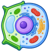
There is no single shape called a human cell. This diagram shows the shared eukaryotic plan before cells specialize for different tissues.
Interactive vector diagram — click a part, hide labels, or download a 300 DPI PNG / editable SVG.
Diagram labels and functions
| Structure / stage | Function |
|---|---|
| Cell membrane | Controls movement into and out of the cell. |
| Cytoplasm | Fluid where many chemical reactions take place. |
| Nucleus | Stores DNA and coordinates cell activity. |
| Mitochondrion | Transfers energy from respiration into ATP. |
| Ribosome | Builds proteins from amino acids. |
| Golgi apparatus | Modifies, sorts and packages proteins. |
A typical human cell
Most human cells have a membrane, cytoplasm, nucleus, mitochondria, ribosomes, ER and Golgi apparatus, but the amounts and shapes vary with function.
Specialized human cells
Neurons extend long axons, muscle fibers pack in contractile proteins, and mature red blood cells lose the nucleus to carry more hemoglobin.
Related diagrams
Frequently Asked Questions
Is a human cell an animal cell?
Yes. Human cells are animal eukaryotic cells.
Do all human cells have a nucleus?
No. Mature red blood cells lose their nuclei, and platelets are cell fragments.
What is the largest human cell?
The egg cell is the largest by diameter.
Search terms covered by this page
human cell diagram