 celldiagram.world
Biology · Cell Structure
Animal Cell Diagram
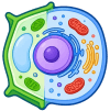
A fully labeled animal cell diagram, drawn as a fixed vector and checked against standard biology curricula. Click any organelle to read what it does, switch the labels off to test yourself, or download a clean copy for homework and worksheets.
Skip to the diagramFixed vector diagram, drawn and checked against standard biology curricula — not AI-generated on the fly.
Cell Parts and Their Functions
| Structure | Function |
|---|---|
| Controls what moves in and out of the cell | |
| Stores DNA and controls cell activity | |
| Releases energy (ATP) from nutrients | |
| Builds and transports proteins and lipids | |
| Packages and ships proteins | |
| Builds proteins from amino acids | |
| Breaks down waste and worn-out parts |
Click a row to highlight that structure on the diagram.
How to read the animal cell diagram
An animal cell is a eukaryotic cell: its genetic material is packed inside a nucleus rather than floating loose in the cytoplasm. The whole cell is wrapped in a partially permeable cell membrane, and everything between that membrane and the nucleus is cytoplasm — a jelly-like fluid in which the organelles sit and move.
Because organelles differ hugely in size, no textbook diagram of an animal cell is drawn to scale. Ribosomes are only about 20 nanometres across, while the nucleus can be 6 micrometres wide, so ribosomes are exaggerated in every labeled diagram of a cell to keep them visible. Treat the picture as a map of what is present and what it does, not as a measurement.
The key organelles and what they do
The nucleus is the control centre: it stores DNA and sends instructions out to the cytoplasm, and the nucleolus inside it builds ribosomes. Those ribosomes assemble proteins, either free in the cytoplasm or attached to the rough endoplasmic reticulum. Finished proteins travel to the Golgi apparatus, which modifies, sorts and packages them into vesicles.
Mitochondria supply the energy for all of this. They carry out aerobic respiration, breaking glucose down to release ATP, which is why cells that work hard — muscle and nerve cells especially — contain thousands of them. Lysosomes handle the clean-up, using digestive enzymes to break down waste and worn-out organelles.
How an animal cell differs from a plant cell
The fastest way to tell the two apart on a diagram is to look for three things. An animal cell has no cell wall, so its outline is soft and irregular rather than box-shaped. It has no chloroplasts, so it cannot photosynthesise and must take in food. And its vacuoles are small and temporary, instead of one permanent central vacuole filling most of the cell.
Everything else on this diagram — nucleus, mitochondria, ribosomes, endoplasmic reticulum, Golgi apparatus, cell membrane and cytoplasm — is found in plant cells too, which is why exam questions usually ask for the differences rather than the similarities.
Common mistakes in exams
Three errors come up again and again. Labelling the outer line of an animal cell as a 'cell wall' — animal cells do not have one. Calling the nucleolus the nucleus, when the nucleolus is the small dark region inside it. And writing that mitochondria 'make' energy: they release energy that is already stored in glucose and transfer it to ATP.
When you label a blank diagram, draw your leader lines so they touch the structure itself and write the name horizontally at the end of the line. Markers usually accept a line that clearly points at the right structure, even if your drawing is rough.
Test Yourself
Three quick questions. No sign-up, no score saved anywhere.
Question 1 of 4 · Score 0
Which organelle is known as the 'powerhouse of the cell'?
Frequently Asked Questions
What is a labeled diagram of an animal cell used for?
It is used to identify each organelle by name and function — the standard format for homework, revision and exam questions on cell structure.
What are the main parts on an animal cell diagram?
Cell membrane, cytoplasm, nucleus with nucleolus, mitochondria, ribosomes, rough and smooth endoplasmic reticulum, Golgi apparatus and lysosomes.
What is the difference between an animal cell diagram and a plant cell diagram?
An animal cell has no cell wall, no chloroplasts and only small temporary vacuoles. A plant cell has a rigid cellulose wall, chloroplasts and one large central vacuole.
Is this animal cell diagram to scale?
No. Like every textbook diagram, organelle sizes are exaggerated so that small structures such as ribosomes stay visible and can be labelled.
Can I download a blank animal cell diagram for a worksheet?
Yes. Switch the diagram to Blank, then use Download PNG to save an unlabeled copy. It is free to print and use in class, with no sign-up.
Which organelle is the powerhouse of the cell?
The mitochondrion. It carries out respiration and transfers the energy released from glucose into ATP, the form cells can actually use.